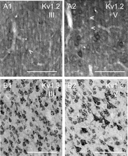
Figure 3. Immunohistochemical expression of Kv1.2 and Kv1.3.
The monoclonal Kv1.2 antibody (A) was obtained from Upstate (Waltham, MA, USA). The monoclonal Kv1.3 antibody (B) was a gift from Dr J. Trimmer. Scale bars = 100 μm. A, monoclonal antibody to Kv1.2. A1, layer III of somatosensory cortex (20×), showing relatively homogeneous staining of neuropil, as well as apical dendrites and perisomatic staining (arrowhead). A2, layer V (20×) showing staining of apical dendrites of pyramidal cells (note arrowheads), as well as perisomatic staining of pyramidal cells. B, monoclonal antibody to Kv1.3. B1, layer III of of somatosensory cortex (20×), showing the grape-like punctate pattern of staining over somas/proximal dendrites. B2, layer V (20×) showing the grape-like cluster pattern of staining over somas/proximal dendrites.

