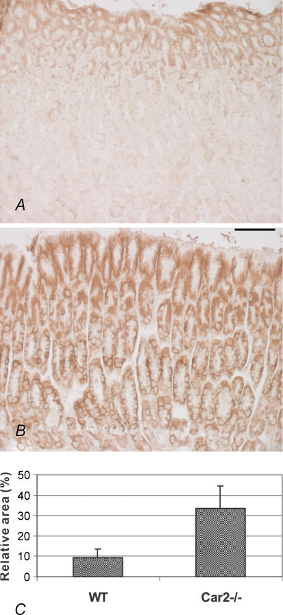
Figure 5. Immunohistochemical staining of CA IX protein in the stomach of the wild-type and CA II-deficient mice.
In the wild-type mouse, the staining is mainly located in the mucus-producing epithelial cells of the outer part of the mucosa (A). In the Car2−/− mouse, the signal was widely spread, covering intensively the whole thickness of the gastric mucosa (B). Original magnifications × 200. C shows digital image analysis results, comparing the staining extent of CA IX protein in the stomach of the wild-type and Car2−/− mice (P = 0.0022). Scale bar, 100 µm, and applies to A and B.

