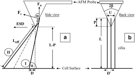
FIGURE 6.
Schematic views of cilia and an AFM probe from two directions. (a) Side view, showing the AFM cantilever and tip aligned with the direction of the ESD. The dot-dashed oblique line is an imaginary continuation of the pyramid side to the cell's surface. Two cilia are also shown (wide lines), to the right and to the left of the dot-dashed line. (b) Three cilia beating in parallel with the ESD into the page and the cantilever of the AFM probe pointing out of the page (toward the reader). The symbols are explained in the text.

