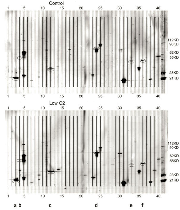
Figure 2.
PowerBlot patterns of JAR cells. Forty elements of various apoptosis-related proteins were spotted onto the membrane. A. Control( exposed to aerobic condition). B. Low O2( exposed to hypoxic condition for 24 hours). After the hypoxia treatment, cell extracts were analyzed with the PowerBlot western array. 40 apoptosis-related proteins described in Table 1 were examined. The membrane was scanned at 700 nm using the Odyssey Infrared Imaging System. *a-f in the Figure are as follows; a:Bad, b:Bag-1, c:caspase-3, d:Hsp70, e:JNK/SAPK, f:PTEN.

