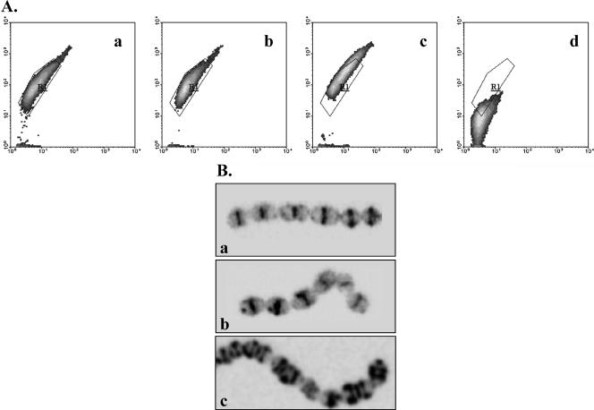
FIG. 5.
(A) Flow cytometric analysis of active cell wall biosynthesis sites of nonadapted (a), acid-adapted (at pH 5.5 for 1 h) (b), acid-habituated (c), and stationary-phase (d) S. macedonicus cells labeled with BODIPY FL vancomycin. Results are presented as density plots of green fluorescence versus side scatter. Region R1 was drawn to enclose the core of nonadapted cells' distribution. (B) CLSM photographs of cells treated as described above (a to c). It should be noted that stationary-phase cells were negative for BODIPY FL vancomycin labeling under CLSM.

