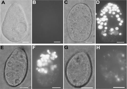
FIG. 5.
Visualizing T4-Tetrahymena interactions by fluorescence microscopy. Tetrahymena cells (35,000/ml) were exposed to SYBR gold nucleic acid stain solution (A and B) or to SYBR gold-labeled T4 phage at 1010 PFU/ml (C and D), 109 PFU/ml (E and F), and 108 PFU/ml (G and H) for 2 h at room temperature. Cultures were fixed in Formalin immediately prior to microscopic analysis. Note that phase-contrast photomicrographs are shown in panels A, C, E, and G; fluorescence photomicrographs are shown in panels B, D, F, and H. Bars are 10 μm in all panels.

