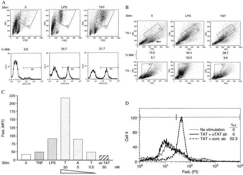
Figure 1.
Tat promotes viability and stimulates human and murine MΦs in culture. (A) Human MΦs. Fluorescence-activated cell sorter (FACscan, Becton Dickinson) analysis of monocytes, enriched from peripheral blood by centrifugal elutriation, cultured for 6 days with either no stimulus (0), 100 ng/ml of LPS, or 50 nM Tat. (Upper) Scatter plot, the large, CD14+ MΦs (M1, 100% CD14+) are gated. FSC, forward scatter. (Lower) Immunofluorescence of all cells in culture after staining with a rhodamine-labeled anti-CD14 antibody (PharMingen). The CD14+ cells (M1, MΦs), as a % of all cells in culture at time of assay, are indicated above, and agree closely with the % MΦs determined by scatter plot. FI, fluorescence intensity. (B) Tat promotes viability of murine MΦs that have been previously activated in vivo. Mouse peritoneal washout cells, a MΦ-rich population, were isolated either 3 days after i.p. TG injection (TG+), or without this in vivo stimulation (TG−). Murine MΦs were cultured for 5 days either in the absence of additional stimulation (0), with LPS (100 ng/ml), or with sTat (100 nM), and then analyzed by flow cytometry scatter plot for a large population of cells (gate, M1, %MΦ) staining for murine MΦ markers (not shown). Arrow indicates population of apoptotic, annexin-reactive cells accumulating in unstimulated cultures. FSC, forward scatter. (C) Median fluorescence (MFl) of monocytes, cultured for 6 days either with no stimulus (0), 50 ng/ml TNF-α, 100 ng/ml LPS, decreasing concentrations of sTat (T, A, T), or 50 nM oxTat (3% H2O2 for 1 hr at 25°C), and stained with an anti-FasL mAb (Nok 1, PharMingen), followed by a fluoresceinated goat anti-mouse polyclonal antibody (Amersham Pharmacia). (D) Neutralization of Tat-mediated induction of FasL on human MΦs by anti-Tat polyclonal antibodies. MΦs cultured for 2 days either with no stimulus, with 50 nM Tat pretreated for 1 hr with murine polyclonal antibodies (1:100 dilution) prepared to oxTat, or with preimmune serum (1:100) were analyzed by FACs after staining with an anti-FasL mAb (Nok 1, PharMingen). FI, fluorescence intensity.

