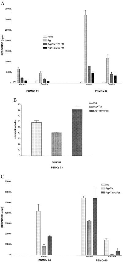
Figure 2.
Participation of FasL in Tat-mediated suppression of lymphocyte proliferation. (A) Human PBMCs from two individuals (PBMCs #1 and #2) cultured in triplicate for 6 days in medium alone, tetanus (0.3 Lf/ml) antigen (Ag) or Candida (4 μg/ml) Ag, or Ags with additionally 125 or 250 nM recombinant Tat protein, were pulsed with [3H]thymidine over the last 18 hr before harvest. Results are representative of 10 experiments with different PBMCs. (B) Human PBMCs from one individual (PBMCs #3) cultured in triplicate for 5 days in either medium (not shown), tetanus antigen (Ag, 0.3 Lf/ml), antigen with the further addition of 50 nM Tat (Ag+Tat), or Ag with 50 nM Tat and recombinant sFas protein (25 μg/ml) to block surface FasL (Ag+Tat+sFas). [3H]thymidine was added over the last 18 hr, and results are graphed as stimulation index (mean cpm stimulated culture/mean cpm medium control). Results are representative of three experiments. (C) Proliferation of PBMCs cultured 6 days with either tetanus (PBMCs #4 and #5) or Candida (PBMCs #5) antigen (Ag) alone as in A, compared with cultures in which Tat (Ag+Tat, 125 nM), or Tat (125 nM) and the antagonistic anti-Fas antibody, ZB4 (250 μg/ml, Upstate Biotechnology) also were added (Ag+Tat+αFas). Results are representative of three experiments.

