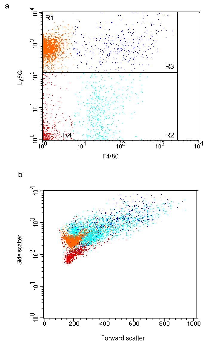
Supplementary Figure 1. Ly-6G+F4/80+ in peritoneal exudates are PMN-macrophage conjugates.

Leukocytes recovered from peritoneal exudates 48 h after peritonitis initiation were stained with anti-Ly-6G and anti-F4/80 and analyzed by flow cytometry. Dot plots represent Ly-6G and F4/80 staining (a) and forward versus side scatter (b). PMNs (R1, orange cells), macrophages (R2, cyan cells) and PMN-macrophage conjugates (R3, blue cells) are indicated. Results are respresentative of six experiments.
