
Fig. 10.
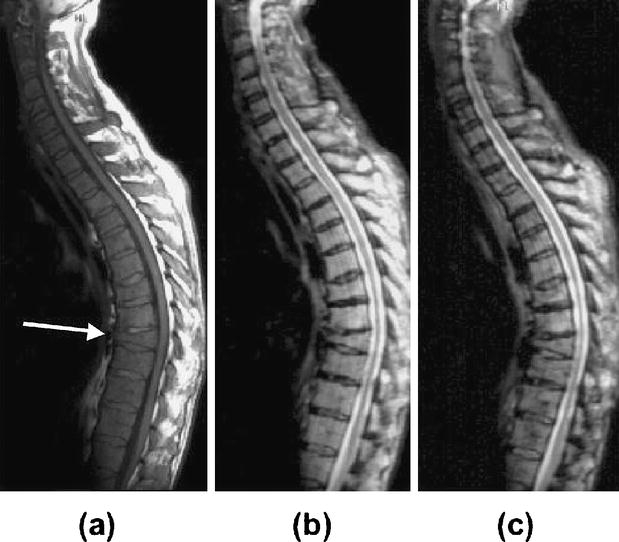
A 57-year-old patient with myeloma after chemotherapy and GCSF treatment. T1-weighted images before (a) show a pathologic fracture of Th 9 (arrow) and a diffuse hypointense signal intensity of the bone marrow in all vertebrae, compatible with a high bone marrow cellularity. Unenhanced STIR images (b) show a diffuse hyperintense bone marrow, also compatible with high bone marrow cellularity. After iron oxide infusion, the hypercellular bone marrow shows only minimal changes in signal intensity on STIR images (c), indicative of a diffuse tumor infiltration. Iliac crest biopsy revealed 80% tumor cells in the bone marrow (figure from [31])