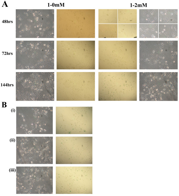
Figure 5.
Prolonged exposure to thymidine results in cellular senescence. (A) Following a 1 mM thymidine 24 hour pre-treatment, cells were electroporated and released into complete medium (1-0 mM) or medium supplemented with 2 mM thymidine (1–2 mM). At 48, 72 and 144 hours, cells were stained for the senescent marker β-galactosidase. The four quadrants for the 1–2 mM sample at 48 hours are combined images obtained from four different fields of view, due to the low confluency of the sample. (B) Images depict varying views and different wells of the 1–2 mM thymidine treated cells at 144 hours. All images are at 20× magnification and counts of individual cells in a representative field were performed to evaluate the degree of senescence induction.

