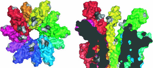
FIG. 8.
Mapping of YscF residues A27, D28, A30, N31, and N47 onto the surface of a T3SS needle. (Left) Surface representation of the end-on view of the Shigella T3SS needle, with each subunit colored differently. The equivalent residues (T23, Q24, L26, Q27, and N43) of the Shigella subunit protein MxiH have been highlighted in gray. (Right) Cutaway surface representation of the side-on view of the Shigella T3SS, with the equivalent Yersinia YscF residues that were mutated highlighted in gray. Class I mutation equivalents A27, A30, and N31 are colored light gray, and class II mutation equivalents D28 and N47 are colored dark gray. This figure was prepared using the PyMOL program (11).

