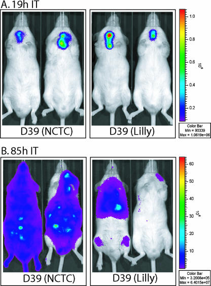
FIG. 5.
Biophotonic imaging of ICR male mice (ca. 25 to 30 g) infected with D39 (NCTC) luxABCDE or D39 (Lilly) luxABCDE. Each mouse was infected intratracheally (IT) with 50 μl of buffered saline containing ∼2.5 × 10⁶ CFU of each strain (see Materials and Methods) (96). Bioluminescence was monitored and recorded at various times in separate animals (87, 88). Typical images from 1-min exposures are shown 19 h (A) and 85 h (B) after infection. The mouse in the lower right corner has cleared the D39 (Lilly) infection.

