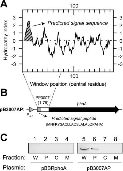
FIG. 2.
Subcellular localization of P. putida DOT-T1E PP3007. (A) Hydrophobicity (Kyte-Doolittle) plot of the PP3007 protein showing the putative signal peptide (window size, 9). The average hydrophobicity of the amino acids included in the window is plotted in the midpoint of the window. (B) Schematic view of the PP3007′-′phoA fusion under the control of the Plac promoter in plasmid pB3007AP (positive for alkaline phosphatase activity). (C) Immunoblot detection of PP3007′-′PhoA in P. putida DOT-T1E(pP3007AP) cell fractions (lanes 5 to 8). P. putida DOT-T1E(pBBRphoA) was used as a negative control (lanes 1 to 4). Protein samples were subjected to electrophoresis (12.5% [wt/vol] SDS-PAGE), followed by Western blotting with an anti-PhoA antibody. Lanes 1 and 5, whole-cell lysate (W); lanes 2 and 6, periplasmic fraction (P); lanes 3 and 7, cytoplasmic fraction (C); and lanes 4 and 8, membrane fraction (M). The positions of molecular mass markers are indicated (in kilodaltons) on the left.

