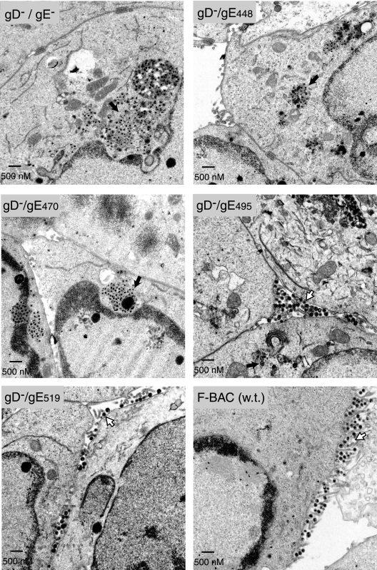
FIG. 2.
Electron micrographs of cells infected with F-BAC gD−/gE CT mutants. Human HEC-1A epithelial cells were infected with F-BAC gD−/gE−, F-BAC gD−/gE448, F-BAC gD−/gE470, F-BAC gD−/gE495, F-BAC gD−/gE519, or F-BAC (w.t.) for 16 h. The cells were fixed and processed for electron microscopy. Black arrows point to aggregates of unenveloped capsids, while white arrows point to enveloped virions on the surfaces of cells.

