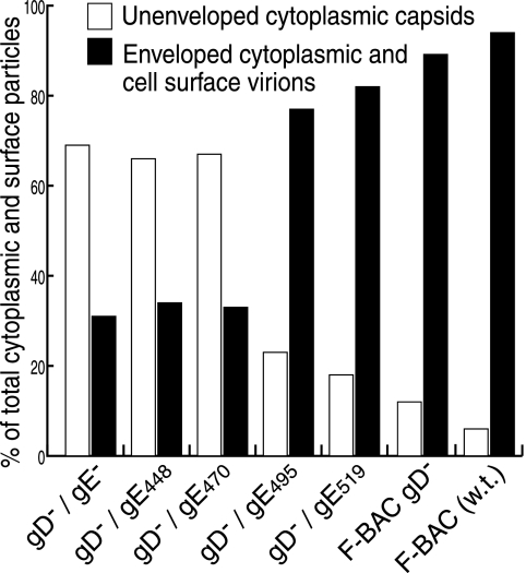
FIG. 3.
Distributions of virus particles in cells infected by gD−/gE CT mutants. Randomly selected sections of HSV-infected HEC-1A cells were characterized by electron microscopy, and unenveloped nucleocapsids in the nucleus and cytoplasm and enveloped virions in the perinuclear space, in the cytoplasm, and on the cell surface were counted. In the figure, values for unenveloped capsids in the cytoplasm (white bars) are compared to those for enveloped virions in the cytoplasm combined with virions on cell surfaces (black bars).

