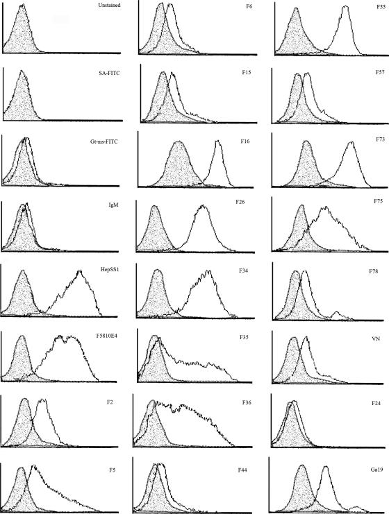
FIG. 3.
Peptide binding to Vero cells treated with GAG lyases. Vero cells (5 × 10⁵ per sample) were treated in suspension for 2.5 h at 37°C without enzymes or in the presence of 4 U chondroitinase C (Sigma) or a mixture of 4 U heparin lyase I (Sigma) and 4 U heparin lyase III (Seikagaku, Japan) in GAG lyase buffer. Cells were then incubated with 10 μg/ml biotinylated peptide or HepSS1 or F5810E4 anti-heparin sulfate antibody for 1 h at 4°C, followed by three washes with FACS buffer. Goat anti-mouse (IgG, IgA, and IgM)-FITC at 1:20 or streptavidin-FITC at 1:100 was added to peptide cell mixtures for 1 h at 4°C in the dark to detect binding of anti-heparin sulfate monoclonal antibodies or biotinylated peptides, respectively, and were used in the absence of antibodies and peptides to measure background fluorescence due to nonspecific binding of the FITC conjugate. Samples were analyzed for staining on a Becton-Dickinson LSR flow cytometer. Unshaded histogram, binding to untreated cells; shaded histogram, binding after heparin lyase I plus heparin lyase III treatment.

