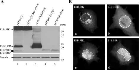
FIG. 2.
Expression and subcellular localization of E1B proteins. (A) H1299 cells were transfected with different constructs (2 μg plasmid) expressing E1B-55K, E1B-156R, E1B-93R, and E1B-84R as indicated at the top. pE1B-55K ΔSD #1415 in contrast to pE1B-55K does not express E1B-156R, E1B-93R, or E1B-84R but only E1B-55K. A Western blot assay of β-actin was included as a loading control. Molecular mass markers are indicated at the right. (B) The subcellular localization of the four E1B proteins was investigated by indirect immunofluorescence analysis of transfected H1299 cells (5 μg of pE1B-55K ΔSD #1415, pE1B-156R #1326, pE1B-93R #1327, or pE1B-84R #1641). Boundaries of nuclei are marked with dashed lines. Magnification, ×7,600.

