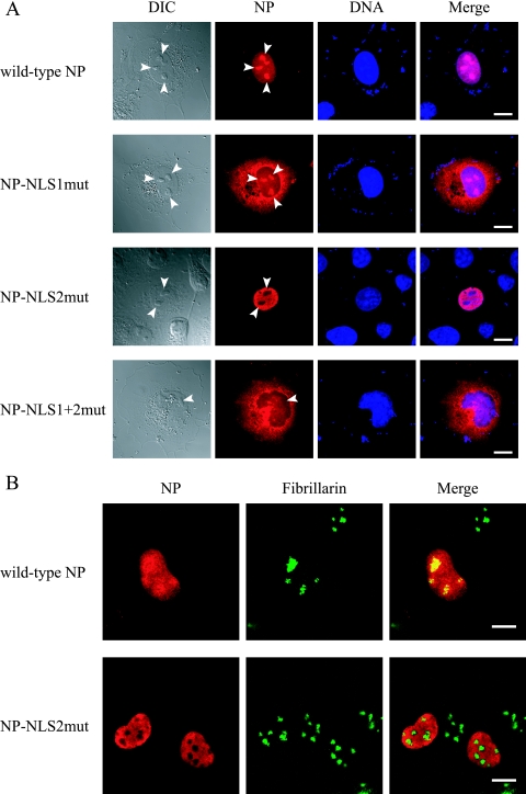
FIG. 2.
Alteration of the subcellular/subnuclear localization of NP following alanine replacements in its NLSs. (A) Subcellular localization of mutant NPs. Plasmids for the expression of wild-type or mutant NP were transfected into COS-7 cells. At 12 h posttransfection, the cells were fixed and incubated with an anti-NP antibody (red) and with Hoechst dye (blue). The far left column shows differential interference contrast images. The far right column shows merged fluorescent images (Merge). Arrowheads point to nucleoli. Scale bars, 10 μm. (B) Subnuclear localization of wild-type NP and NP-NLS2mut. Plasmids for the expression of c-Myc-tagged wild-type NP or NP-NLS2mut were transfected into COS-7 cells. At 12 h posttransfection, cells were fixed and incubated with an anti-c-Myc antibody (red) and with an antibody against the nucleolar protein fibrillarin (green). The right column shows merged fluorescent images.

