FIG. 3.
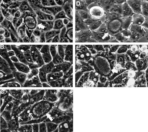
Phase-contrast microscopic detection of doubly resistant recombinant inclusions at the first passage (P-1) of IFU produced in cells that were simultaneously infected with two kinds of antibiotic-resistant mutants. (A) A normal 48-h p.i. inclusion (thick arrow) formed in the absence of antibiotics used to select for recombinants. Inclusions formed by each strain of C. trachomatis used in this study would have similar appearances in antibiotic-free culture medium or in a medium containing just that antibiotic to which the strain was resistant. (B) A typical 46-h p.i. field in a cross no. 1 (OFXr-1 × LINr-1) set A P-1 flask derived from a primary (P-0) flask in which all cells were infected with only parental strain OFXr-1. The P-1 flask contained OFX plus LIN. OFXr-1 is sensitive to LIN and formed tiny, inhibited inclusions in some cells. Similarly, LINr-1, the other parental strain in cross no. 1, is sensitive to OFX and would also be unable to form normal inclusions in OFX plus LIN medium (not shown). (C) A 48-h p.i. field in a cross no. 1 set C P-1 flask derived from a P-0 flask in which all cells were simultaneously infected with OFXr-1 and LINr-1. The P-1 flask contained OFX plus LIN medium, in which parental IFU formed inhibited inclusions (small arrows) and OFXr LINr recombinants formed normal inclusions (large arrow). (D) A typical 68-h p.i. field in a cross no. 3 set C P-1 flask derived from a P-0 flask in which all cells had been simultaneously infected with OFXr-1 and RIFr-1. The P-1 flask contained OFX plus RIF medium. An OFXr RIFr recombinant inclusion is shown (large arrow) along with numerous inhibited inclusions (small arrow) formed by nonrecombinant parental IFU. (E) A typical 46-h p.i. field in a cross no. 4 set C P-1 flask derived from a P-0 flask in which all cells were simultaneously infected with OFXr-1 and TMPr-1. The P-1 flask contained medium supplemented with OFX and TMP. An OFXr TMPr recombinant inclusion is shown (large arrow). Also shown are examples of relatively large, empty-appearing inclusions (small arrow) that are typically formed by TMP-sensitive strains of C. trachomatis (OFXr-1 in this experiment) in the presence of TMP. Spontaneous mutants of parental strains would also be able to form doubly resistant inclusions in P-1 flasks containing two antibiotics, but their rarity (<10−7) would prevent their detection by microscopic scanning in practice.

