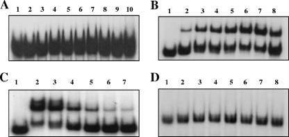
FIG. 4.
EMSA analysis of meningococcal Fur binding to meningococcal secY and nspA operator DNA probes. DNA probes used are as follows: A, secY; B and C, nspA; D, rmp. One nanogram of 32P-labeled DNA fragments was incubated with increasing concentrations of N. meningitidis Fur protein. (A) Lane 1, no Fur; lanes 2 to 10, 80 nM, 120 nM, 140 nM, 160 nM, 180 nM, 200 nM, 240 nM, 300 nM, and 400 nM Fur, respectively. (B and D) Lane 1, no Fur; lanes 2 to 8, 160 nM, 320 nM, 400 nM, 480 nM, 560 nM, 720 nM, and 800 nM Fur, respectively. (C) EMSA analysis of 32P-labeled nspA DNA after incubation with meningococcal Fur and an excess of cold probe (unlabeled operator DNA fragments). Lane 1, no Fur and no cold probe; lane 2, 800 nM Fur and no cold probe; lanes 3 to 7 800 nM Fur and 50-fold, 100-fold, 200-fold, 300-fold, and 400-fold cold probe, respectively.

