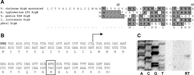
FIG. 2.
Determination of transcriptional and translational start sites for V. cholerae higB. (A) Alignment of the 5′ ends of annotated V. cholerae HigB and characterized or putative HigBs from Salmonella enterica serovar Typhimurium, Yersinia pestis, Idiomarina loihiensis, and Rts1. Conserved amino acids are shaded. (B) Annotated and experimentally determined start sites for V. cholerae higB and HigB. The annotated translational start is shown in bold italics, the experimentally determined translational start site is boxed, and the experimentally determined transcriptional start site is underlined and marked with a bent arrow. (C) Primer extension analysis of the 5′ end of V. cholerae higB transcripts. The sequence corresponds to the bottom strand shown in panel B. RT, reverse transcription reaction performed with a higB-specific primer. The same start site was also identified using 5′ rapid amplification of cDNA ends (data not shown).

