FIG. 3.
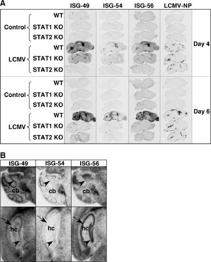
Anatomic localization of ISG-49, ISG-54, ISG-56, and LCMV NP RNA in the brain. WT, STAT1 KO, and STAT2 KO mice were injected i.c. as described in the legend to Fig. 1, and brains were removed at days 4 and 6 for in situ hybridization. Paraffin-embedded sagittal sections (8 μm) were hybridized with 33P-labeled cRNA probes transcribed from linearized ISG-49, ISG-54, ISG-56, or LCMV NP RNA plasmids and exposed to Kodak MR film for 3 days as outlined in Materials and Methods. (A) No hybridization above background of the ISG-49, ISG-54, ISG-56, or LCMV NP probes was evident in uninfected (control) brains. Following LCMV infection, in WT mice high levels of ISG-49 and ISG-56 hybridization were evident throughout the brain at day 4 and increased further by day 6. In contrast, there was a lower and more restricted level of ISG-54 hybridization. Increased ISG-49 and ISG-56 hybridization was also evident in the brain of STAT1 KO mice at day 4 but not day 6 postinfection, particularly in the meninges and ventricles. A small increase in ISG-49 hybridization was evident in the meninges and choroid plexus in the brain of STAT2 KO mice at day 6 postinfection. (B) Differential patterns of ISG-49, ISG-54, and ISG-56 hybridization at day 6 postinfection. In the Purkinje cell layer (arrowhead) of the cerebellum (cb), high levels of ISG-49 but low levels of ISG-54 and ISG-56 hybridization were seen. In the CA1 pyramidal neurons (arrow) and dentate gyrus (arrowhead) of the hippocampus (hc), high levels of ISG-56 but low levels of ISG-49 and ISG-54 hybridization were seen.

