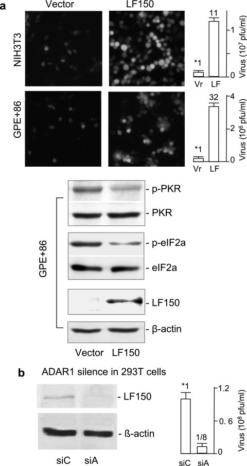
FIG. 4.
ADAR1 mediates host susceptibility to VSV infection. (a) ADAR1 expression. NIH 3T3 (top) or GP+E86 (bottom) cells stably expressing LF150 or the vector were infected with the recombinant VSV-EGFP1 (5). NIH 3T3 cells were infected at an MOI of 10 PFU/cell for 20 min and GP+E86 cells at an MOI of 100 PFU/cell for 1 h. After 10 to 12 h of infection, the infected cells were monitored under fluorescence microscopy (left). The virus titer in the culture medium was quantitatively determined by the PFU (right; n = 4). The number over the bar indicates relative susceptibility to VSV infection. Levels of LF150, total PKR, and eIF-2α, and the phosphorylated PKR and eIF-2α in the testing cells, were examined by immunoblotting, and the results are shown below. (b) ADAR1 knockdown. 293T cells were transfected with siRNA against ADAR1 (siA) or a bacterial gene (siC) for 48 h and infected with VSV for 12 h. Endogenous ADAR1 was analyzed by immunoblotting using the antibody against human ADAR1. The virus titers in media were analyzed as described above. The error bars represent standard deviations.

