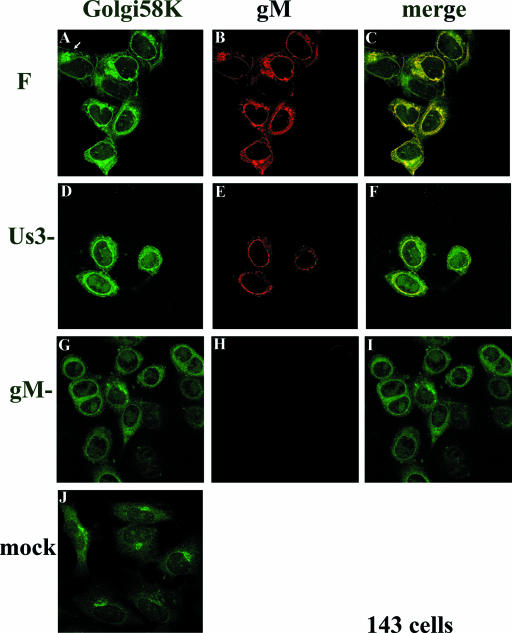
FIG. 3.
Indirect immunofluorescence of infected and mock-infected 143 cells stained with gM or a Golgi-specific marker. 143 cells were infected with the indicated viruses. At 16 h postinfection, the cells were fixed and immunostained as described in the legend to Fig. 2. All images are of single optical sections collected using identical laser and photomultiplier tube (PMT) settings. The arrow in panel A indicates a cell with a Golgi 58K staining pattern that is reminiscent of that of mock-infected cells and different from the staining pattern of other infected cells in the same panel. F, HSV-1(F).

