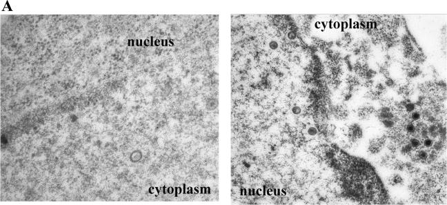
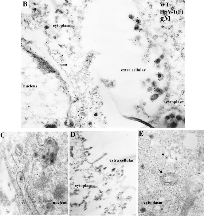
FIG.6.
(A) Immune transmission electron microscopy of HEp-2 cells infected with the gM-null mutant and reacted with gM specific antibody. HEp-2 cells were infected with the gM-null virus R7216, fixed at 14 h after infection, and reacted with anti-gM rabbit polyclonal serum. Bound antibody was visualized by reaction with the F(ab)2 fragments of a goat anti-rabbit antibody conjugated to 12-nm colloidal gold particles. In this and subsequent electron photomicrographs, capsids of approximately 125 nm serve as a size standard. Magnifications: left panel, ×45,000; right panel, ×30,000. (B to E) Electron photomicrograph of a thin section of HEp-2 cells infected with HSV-1 (F) and stained with antibody to gM. HEp-2 cells were infected at 5.0 PFU per cell, harvested at 14 h after infection, fixed, embedded, sectioned, and reacted with rabbit gM-specific antibody and, subsequently, goat anti-rabbit IgG conjugated to 12 nm gold beads as described in Materials and Methods. In panel D, an arrow indicates heavily immunolabeled reduplicated membrane in cytoplasm. Arrowhead indicates a group of immunolabeled virions located within a cytoplasmic vacuole. Magnifications: C, D, and E, ×30,000; B, ×45,000.


