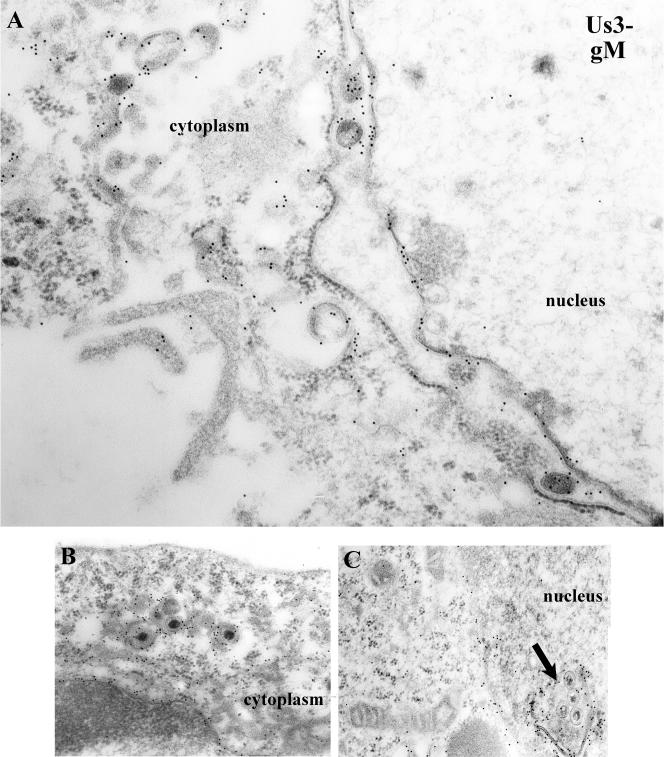
FIG. 7.
Electron photomicrograph of thin sections of HEp-2 cells infected with a US3 mutant of HSV-1(F) and stained with gM-specific antibody. Cells were infected with R7039, a viral mutant lacking US3. At 14 h postinfection, the cells were fixed and embedded. Thin sections were reacted with gM-specific antibody, followed by reaction with 12-nm gold-conjugated anti-rabbit IgG, and examined in a Phillips 201 electron microscope. In panel C, an arrow indicates a cross-section of a punctate extension or invagination of the nuclear membrane containing virions. This feature is peculiar to cells infected with the US3 deletion virus. Magnifications: A, ×45,000; B and C, ×30,000.

