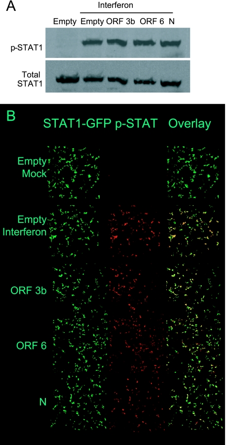
FIG. 8.
Analysis of STAT1 phosphorylation by SARS-CoV proteins. A. 293T cells were transfected with the indicated plasmid for 24 h and then treated with IFN-β for 1 h. Cells were harvested, and lysates were analyzed by Western blot analysis using antibodies recognizing the phospho- and total forms of STAT1. B. Cells were transfected with the indicated plasmid and STAT-1 GFP for 24 h and then treated with IFN-β for 1 h. Cells were fixed, permeabilized, and analyzed for phospho-STAT1 by confocal microscopy using a 10× objective. Representative images are shown.

