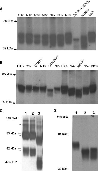
FIG. 4.
Analysis of E2 glycosylation mutants was done by Western immunoblotting. SK6 monolayers were infected (MOI of 1) with each of the mutants or parental BICv or mock infected and harvested 48 h postinfection. Cell lysates were run under reducing (A, B, and D) or nonreducing (C) conditions in 12% sodium dodecyl sulfate-polyacrylamide gels. CSFV E2 was detected with CSFV E2 monoclonal antibody WH303. (C) Lane 1, BICv; lane 2, N1v; lane 3, ΔO1N1-N6/N3v. Asterisks indicate, from top to bottom, E2 homodimers, E1-E2 heterodimers, and monomeric E2, as described by Weiland et al. (44). (D) Lane 1, untreated BICv; lane 2, PNGase F-treated BICv; lane 3, untreated ΔO1N1-N6/N3v.

