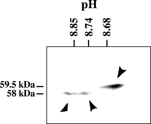
FIG. 4.
2-D gel electrophoresis of the Vhs polypeptides. Vero cells were harvested 24 h after infection with 10 PFU/cell of wild-type HSV-1, and solubilized in 2-D gel sample buffer. The lysate was analyzed by 2-D gel electrophoresis, involving isoelectric focusing in the first dimension and SDS-PAGE in the second. Following electrophoresis, the proteins were transferred to an Immobilon P membrane and detected by immunoblotting using rabbit antiserum raised against a UL41-lacZ fusion protein. The three predominant Vhs polypeptides are indicated by arrowheads. The positions of the 58-kDa and 59.5-kDa polypeptides are indicated to the left of the gel, while the pH values at different positions in the isoelectric focusing gel are indicated at the top.

