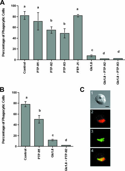
FIG. 5.
PTP-H2 and -H3 significantly reduce the phagocytosis of E. coli and polystyrene beads by S2 cells. Cells were transfected with pIZT/PTP-H1, -H2, -H3, -J1, or the empty vector (Vector) or cotransfected with PTP-H2 or PTP-H3 plus pIZT/Glc1.8. Forty-eight hours posttransfection, rhodamine-conjugated E. coli (A) or fluorescent polystyrene beads (B) were added to cells. Cells were examined 45 min later for phagocytosis by epifluorescence microscopy (n = 6 replicates per treatment). Means with the same letter in panel A or B are not significantly different (P < 0.05) by the Tukey-Kramer multiple-comparison procedure. (C) PTP-H2 colocalizes with Fak in focal adhesions. A light micrograph of a typical S2 cell expressing PTP-H2 is shown in image 1. The same cell after staining with anti-FakY397 (red) indicates that Fak56 localizes primarily to focal adhesions (2). Staining with anti-V5 (green) indicates that PTP-H2 localizes to the same region of the cell (3). Merging of images 2 and 3 results in an orange-yellow signal indicative of colocalization of Fak56 and PTP-H2. The scale bar in panel C1 equals 10 μm.

