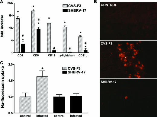
FIG. 4.
Immune cell accumulation in the CNS tissues and BBB permeability in mice infected with SHBRV-17 versus CVS-F3. (A) Levels of mRNAs specific for CD4, CD8, CD19, κ light chain, and CD11b in the cerebellum of the CVS-F3-infected, SHBRV-17-infected, and uninfected mice described in the legend to Fig. 2 were measured by quantitative RT-PCR as detailed in Materials and Methods. The results are expressed as fold increases in mRNA levels in infected versus uninfected mice. Statistically significant differences determined by the Mann-Whitney test between infected versus uninfected mice and between CVS-F3- versus SHBRV-17-infected mice are denoted by the symbols * (P < 0.001) and # (P < 0.05), respectively. (B) Accumulation of CD4-positive cells in the cerebellum of uninfected and CVS-F3- or SHBRV-17-infected mice were identified by immunohistochemistry with phycoerythrin-conjugated anti-mouse CD4 monoclonal antibody as described in Materials and Methods. Photomicrographs are presented at a final magnification of ×200. (C) BBB permeability in the cerebellum was assessed by measuring Na-fluorescein leakage from the circulation into CNS tissues 8 days following infection with CVS-F3 and SHBRV-17 as described in Materials and Methods. The data are expressed as fold increases in Na-fluorescein content over background values obtained with tissues from similarly treated uninfected mice. Statistically significant differences between the groups determined by the Mann-Whitney test are denoted by asterisks (P < 0.05, n = 6).

