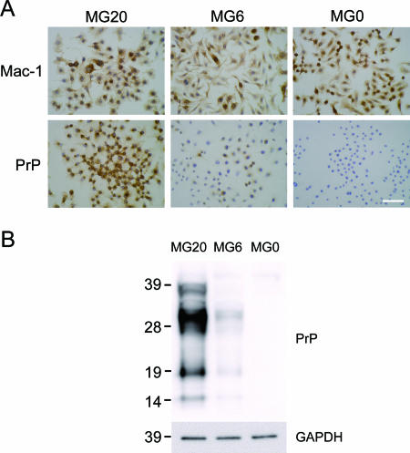
FIG. 1.
Immunostaining and PrP expression of microglial cell lines derived from PrP-overexpressing (MG20), C57BL/6 (MG6), and PrP-deficient (MG0) mice. (A) Each cell line was immunolabeled with anti-Mac-1 and PrP antibodies and stained with 3,3-diaminobenzidine substrate as chromogen (brown). Nuclei were lightly counterstained with hematoxylin (blue). Scale bar, 50 μm. (B) The levels of PrP were compared in microglial cell lines by semiquantitative immunoblotting with MAb T2. Proteins in postnuclear cell extracts prepared without proteinase K treatment were precipitated with acetone prior to electrophoresis. Molecular mass standards (kDa) are indicated on the left. GAPDH (glyceraldehyde-3-phosphate dehydrogenase) was used as a control to confirm protein content of cell extracts.

