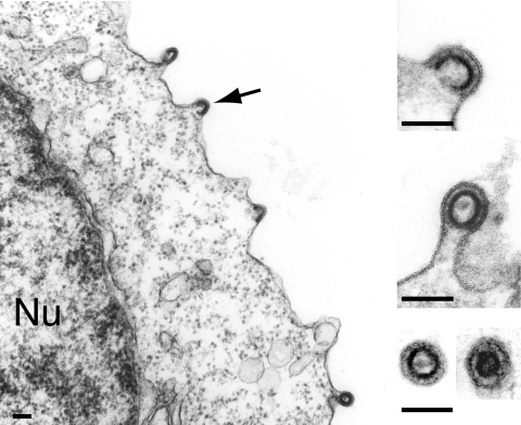
FIG. 3.
Plasma membrane budding and release of MusD particles upon restoration of the Gag myristoylation signal and basic domain. Electron microscopy analysis of human 293T cells transfected with the MusDmyr+basic construct. Same experimental conditions as in Fig. 1B and C. A low-magnification image is shown on the left with budding particles (arrow), and enlarged views are shown on the right, with two free particles (immature and mature) in the cell supernatant (bottom image). Bars, 0.1 μm.

