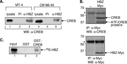
FIG. 2.
HBZ and CREB interact in vivo and in vitro. (A) HBZ and CREB interact in HTLV-1-infected cells. Cell extracts from the infected cell lines MT-4 (lanes 1 to 3) and C8166-45 (lanes 4 to 6) were immunoprecipitated with a preimmune serum (PI) or an HBZ antiserum (α-HBZ). The complexes were separated by SDS-polyacrylamide gel electrophoresis (PAGE), transferred to a nitrocellulose membrane, and probed with an anti-CREB antibody. A fraction of the whole-cell extract is shown in lane 1. (B) HBZ and CREB interact in uninfected cells. Cell extracts from CHOK1-Luc cells transfected with empty vector (−) or an expression plasmid for HBZ tagged with Myc (HBZ-Myc) were immunoprecipitated with an anti-Myc antibody. The complexes were separated by SDS-PAGE, transferred to a nitrocellulose membrane, and probed with an anti-CREB antibody (upper panel) or an anti-Myc antibody (lower panel). Protein standards (in kilodaltons) are indicated at the left. The asterisk indicates the heavy chain of immunoglobulin G. (C) HBZ and CREB interact in vitro. 35S-labeled HBZ was incubated with either GST (10 pmol, lane 2) or GST-CREB (10 pmol, lane 3). Bound HBZ protein was detected by PhosphorImager analysis. A fraction of the input protein (35%) is shown in lane 1.

