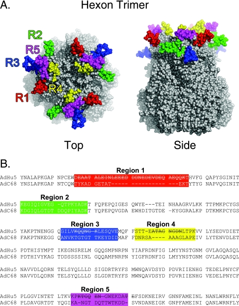
FIG. 1.
Structure and sequence of AdC68 adenovirus hexon. (A) Space-filling representation of the crystal structure of the trimeric AdC68 hexon showing the potential epitope regions (43). Potential epitope regions on hexon are located in the three tower regions at the top of the molecule. These form the exterior surface of the virion. The regions are labeled on the sequence and highlighted in the same color on the molecule: R1 (red), R2 (green), R3 (blue), R4 (yellow), and R5 (magenta). Although R3 is on the upper surface of hexon, it is buried between hexons in the intact virion and so is not accessible to antibodies. The figure was produced with PyMol v0.99. (B) Partial sequence alignment of the 932-residue AdC68 and 951-residue Ad5 hexons based on an alignment of the structures, showing residues 121 to 474 of Ad5 and 121 to 455 of AdC68. Amino acid residues in flexible regions that were not observed in the Ad5 X-ray structure are indicated by lines through the sequence. The potential epitope regions are colored as in panel A.

