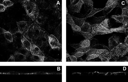
FIG. 1.
VP4 mostly localizes at the apical membranes of Caco-2 cells, whereas no particular localization was observed in MA 104 cells. MA 104 cells (5 days old) and Caco-2 cells (20 days old) were infected with the RF rotavirus strain at 3 PFU/cell and 10 PFU/cell, respectively. VP4 was detected after cell permeabilization, using monoclonal antibody 7.7 and a fluorescein isothiocyanate-labeled secondary anti-mouse IgG antibody, and observed by confocal microscopy as described in Materials and Methods. (A and B) MA 104 cells. (C and D) Caco-2 cells. (A and C) xy projections. (B and D) xz sections.

