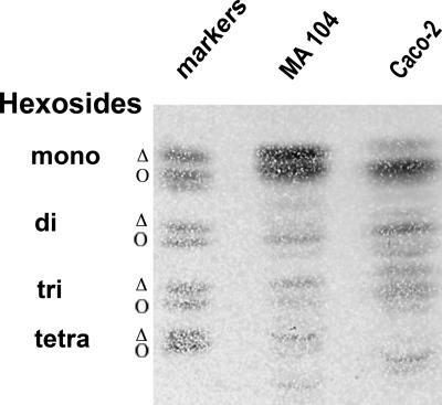
FIG. 6.
Profiles of glycolipids from Caco-2 and MA 104 cells are highly different. Cells were scraped into TNE buffer and sonicated, and glycolipids were extracted as described in Materials and Methods. Glycolipid amounts corresponding to 1 mg protein in MA 104 and Caco-2 cell homogenates were spotted onto silica HPTLC plates. Migration was performed in chloroform-methanol-H2O (65/25/4 [vol/vol/vol]). The plate was sprayed with orcinol for glycolipid visualization. Glycolipid markers were mono (or cerebrosides)-, di (or lactosyl-ceramides)-, tri (or globoside 3)-, and tetra (or globoside 4)-hexosides. For mono-, di-, and trihexosides, two main bands were revealed that corresponded to the nonhydroxylated (Δ) and hydroxylated (Ο) forms of the fatty acid chain esterifying the ceramide moiety.

