FIG. 8.
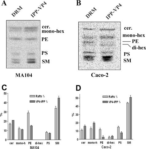
Cell type differences in the lipid compositions of VP4-associated DRM subsets isolated from MA 104 and Caco-2 cells. (A and B) Typical separations by HPTLC of lipid extracts from [14C]serine-labeled MA 104 (A) and Caco-2 (B) cells. Lipids from DRM and immunoprecipitated (IPP) fractions, as described in the legend to Fig. 7, were extracted and separated by HPTLC as described in the legend to Fig. 6. Radioactivity was detected by phosphorimager screening. Migration of lipid standards is indicated. Cer, ceramide; hex, hexoside; PS, phosphatidylserine; PE, phosphatidylethanolamine; SM, sphingomyelin. (C and D) Quantification of the data in panels A and B was performed using ImageQuant software. Data are expressed as the percentage of each lipid species, considering 100% to be the sum of all species in each sample (n = 3).

