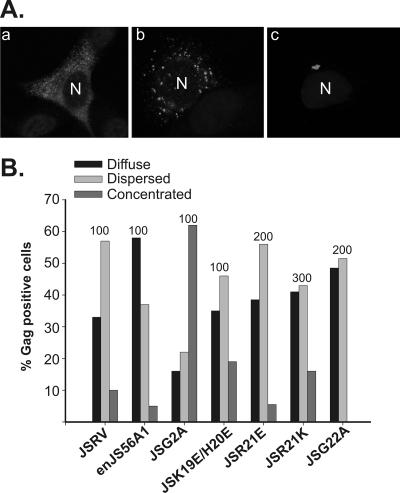
FIG. 2.
Patterns of Gag staining by confocal microscopy of cells expressing JSRV, enJS56A1, and M domain mutants. (A) By confocal microscopy, Gag staining in HeLa cells expressing JSRV and enJS56A1 is described as diffuse (panel a), dispersed (panel b), or concentrated (panel c) (19). The figure represents typical examples of enJS56A1-expressing cells; anti-MA staining is displayed in gray, and the letter “N” indicates the location of the nucleus. (B) Gag staining patterns of JSRV and enJS56A1 M domain mutants in a representative experiment. The number on top of each bar indicates the total number of cells counted for each virus/mutant.

