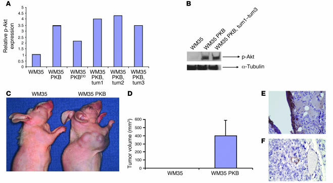
Figure 2. Akt causes vertical growth melanoma in vivo.
(A) Parental WM35 cells, pooled Akt-transduced WM35 cells prior to implantation in vivo (WM35 PKB and WM35 PKBDD), and cells derived from tumors of mice injected with WM35 PKB cells (tum1–tum3) were examined for expression of phosphorylated Akt (p-Akt). The experiment was performed in triplicate. (B) Representative Western blot of phosphorylated Akt and α-tubulin; overexpression converted radial growth melanoma to vertical growth melanoma. (C and D) Mice (n = 4 per group) were injected with either vector control or Akt-expressing melanoma cells and were observed at 1 month. (E) Immunohistochemistry for VEGF. A high level of VEGF expression was noted, especially surrounding necrotic areas. (F) Smooth muscle actin immunohistochemistry, demonstrating that tumors were highly angiogenic. Note the invasion of tumor cells into vessel at the image’s center. Original magnification, ×100.

