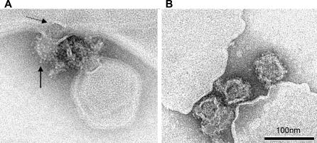
Figure 7. Electron Micrographs of TBE Virus Interacting with Liposomes at pH 5.4 and 10.0.
(A) Electron micrographs of a virus particle in the process of low pH–induced fusion with a liposome. Solid arrow points to low pH–induced projections at the virion surface; dotted arrow points to an electrodense structure presumed to be the nucleocapsid in the process of release.
(B) Virus particles attached to liposomal membranes at alkaline pH. Negative stain by phosphotungstic acid adjusted to pH 8.0.

