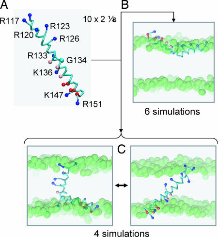
Fig. 2.
Interactions of the S4 helix with a PC bilayer. (A) CG model of the S4 helix (residues 115–153) from KvAP showing the location of the side chain particles for basic (blue), acidic (red), and polar (pink) residues. This model, in a box of 256 DPPC molecules and 3,150 water particles, was the starting point for five CG-MD simulations, each of duration 2 μs. (B) Snapshots from the end of the S4 CG-MD simulations showing the S4 helix located at the lipid/water interface. (C) Snapshots from the end of the S4 CG-MD simulations showing the S4 helix inserted in the bilayer and switching between an extended helix and kinked helix conformation. The glycerol backbone particles are shown as green spheres.

