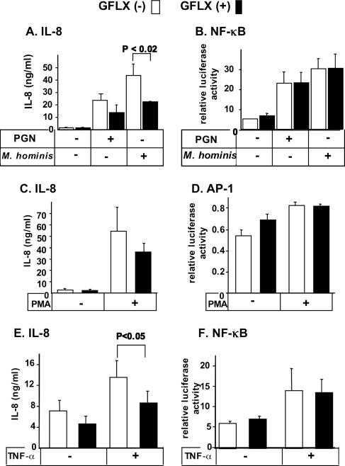
FIG. 2.
GFLX suppresses stimulated IL-8 secretion from PC-3 cells but fails to alter the activation of NF-κB and AP-1. PC-3 cells were plated at 5 × 10⁴ cells per well in 24-well plates. After 24 h of incubation, the cells were transiently transfected by use of the FuGENE 6 transfection reagent with 179.1 ng of an NF-κB or an AP-1 reporter construct and 20.8 ng of pRL-TK. GFLX was added at 16 μg/ml after 24 h of incubation. Forty-eight hours after transfection, the cells were stimulated with 1 μg/ml PGN (A and B), 0.1 μg/ml M. hominis (A and B), or 50 μg/ml PMA (C and D) for 6 h or with 10 ng/ml TNF-α (E and F) for 2 h. The concentrations of IL-8 secreted into the medium (A, C, and E) were determined by an enzyme-linked immunosorbent assay, and the luciferase activities for NF-κB (B and F) and AP-1 (D) were measured by the dual luciferase reporter assay described in Materials and Methods. Open and closed bars, incubation without and with GFLX, respectively. The data shown are the means ± SDs for three experiments. P < 0.02 and P < 0.05 indicate the P values for the results for incubation with GFLX compared with the results for incubation without GFLX.

