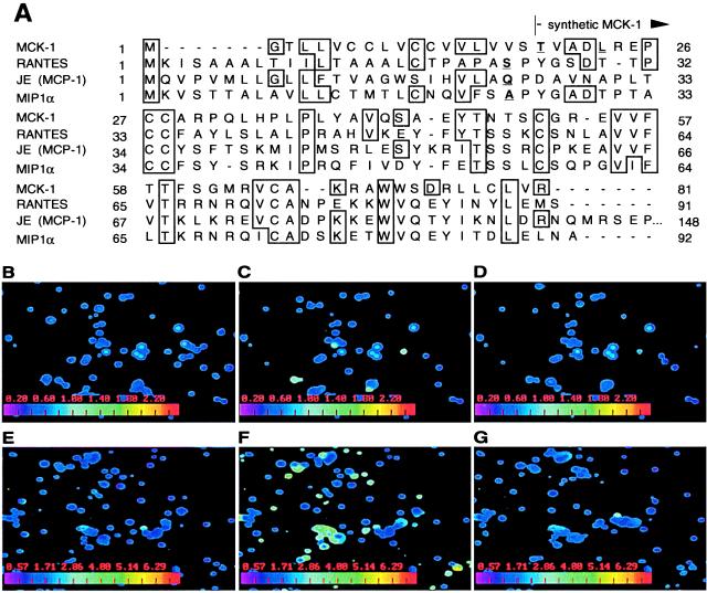
Figure 1.
MCK-1 induces Ca2+ signaling in murine PECs. (A). Predicted MCK-1 amino acid sequence (3) showing the similarity of this protein to three murine β chemokines, RANTES, JE (the homolog of human MCP-1), and MIP1α. The predicted signal sequence cleavage sites of the displayed chemokines are indicated in blue, including MCK-1 threonine 19, at which the synthetic MCK-1 peptide commences. (B–G). Digital fluorescence video microscopy frames of synthetic MCK-1 induced Ca2+ flux in glass-adherent PECs. Cells harvested by lavage from uninfected (B–D) or RM427+-infected (E–G) 10-week-old BALB/c mice were labeled with 1 μM Fura-2-AM, and levels of free intracellular Ca2+ in individual cells, indicated on the digitally calculated color scale inserts on each panel, were measured by spectrofluorometric video microscopy before (B and E), 12 sec after addition of 250 nM MCK-1 (C and F), and 1 (D) and 2 (G) min later. The full series E-G can be viewed at http://cmgm.stanford.edu/∼saederup/

