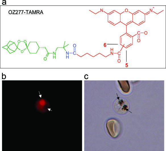
FIG. 4.
Immunofluorescence labeling of trophozoites. (a) Chemical structure of RBX11160-TAMRA. (b) Immunofluorescent staining of two parasites in a single erythrocyte by labeled RBX11160 (a). One trophozoite shows intense staining of the food vacuole (indicated by a long white arrow), whereas the other shows negative staining of the pigment-containing food vacuole (short white arrow) with diffuse cytosolic staining. (c) Bright-field view of panel b. Black arrows indicate parasite pigments in the food vacuole.

