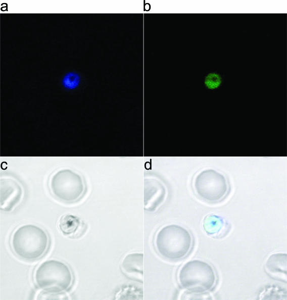
FIG. 5.
Confocal imaging of trophozoite stage. (a) Trophozoite labeled with ER-Tracker Blue showing cytosolic staining with negative staining of the food vacuole. (b) Corresponding bright-field image of panel a. (c) Parasite from panel a showing labeling with RBX11160-TAMRA. (d) Merged images (panels a to c).

