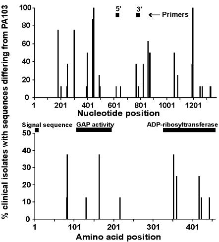
FIG. 3.
Position and frequency of exoT DNA and amino acid sequence variations (n = 8) and the position of PCR primers and signal sequence, GAP, and ADP-ribosyltransferase domains. The x axis represents the nucleotide or amino acid position; the y axis represents the percentage of P. aeruginosa isolates that differed in sequence from PAO1 at each position.

