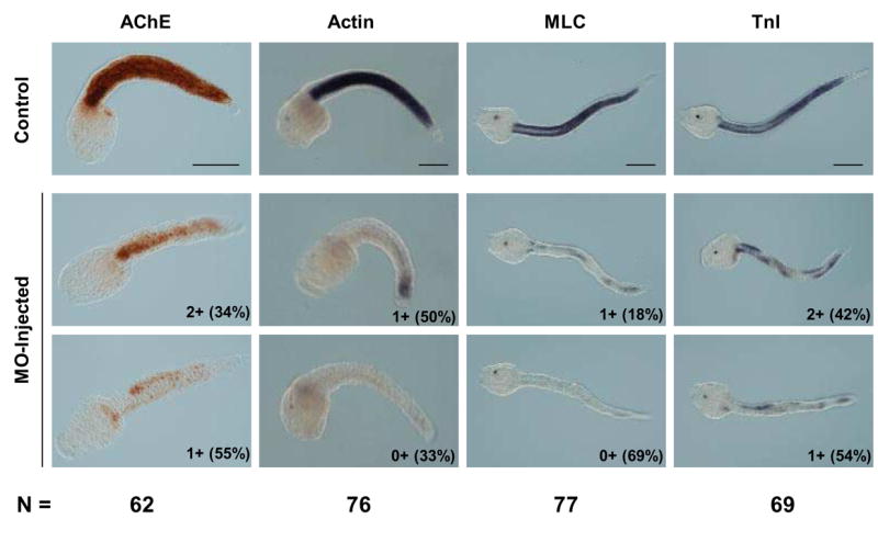
Fig. 5.

MOs targeting Ci-MRF mRNA reduce muscle gene expression in tail formation stage embryos. AChE histochemistry and Actin in situ hybridization were carried out at the mid tail formation stage; MLC and TnI in situ hybridizations were done at the late tail formation stage. Numbers outside of parentheses in the lower right corner of each MO-injected embryo refer to the stain intensity relative to the stain intensity of control embryos reacted for the same time. The system used for classifying embryos (e.g. 3+, 2+, 1+, 0+) is described in the text. Examples are shown of the two most abundant embryo classes for each marker tested and the percentage of embryos in that class is shown in parentheses. The total number of MO-injected embryos analyzed for each marker (N) is shown at the bottom of the figure. Scale bars = 100 μm.
