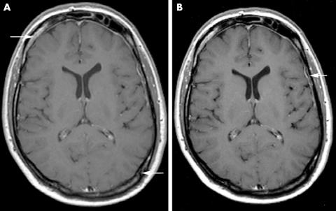
Figure 1 (A) T1 weighted axial MR scan of a 43 year old male patient after contrast application shows meningeal inflammatory involvement with 3 mm thickening of the left hemispherical and right frontal meninges (arrows). (B) Six months after treatment with infliximab (5 mg/kg body wt) at weeks 0, 2, 6 and 8‐weekly thereafter, meningeal contrast enhancement and thickening had clearly regressed.

An official website of the United States government
Here's how you know
Official websites use .gov
A
.gov website belongs to an official
government organization in the United States.
Secure .gov websites use HTTPS
A lock (
) or https:// means you've safely
connected to the .gov website. Share sensitive
information only on official, secure websites.
